Is Muscle an Organ? What Skeletal Muscle Does for Metabolism, Glucose and Aging
Anatomically the answer is yes, and it is a more interesting yes than it first appears…

By
September 11, 2026

Is muscle an organ? Anatomically the answer is yes, and it is a more interesting yes than it first appears. Each whole skeletal muscle is an organ in its own right, built from muscle fibers together with connective tissue, nerve tissue and blood vessels, and the human body holds more than 600 of them.
Collectively they also behave as a distributed metabolic and endocrine organ. Muscle secretes signaling molecules into the blood, it takes up much of the glucose you absorb after a meal, and it runs the internal signaling pathways that adaptation depends on.
That is the underrated part. Muscle is usually taught as the tissue that moves you around, when it is also one of the body’s largest regulators of metabolic health, and this article covers all three of those routes.
What Muscle Is and What It Does
Muscle is an excitable tissue that converts chemical energy, stored as ATP, into mechanical energy. That conversion is the foundation everything else in muscle physiology is built on, from cross-bridge cycling to the interaction between actin and myosin.
Excitable means the tissue responds to a stimulus with a change in the electrical charge across its membrane. All three muscle types are excitable in that sense, though they do not all do it the same way. Skeletal and cardiac muscle normally generate a full action potential, while smooth muscle may use an action potential, a graded change in membrane potential, or signaling pathways that alter contraction without an action potential at all.
What differs between them is where the triggering signal comes from, and that turns out to be the cleanest way to tell them apart.
The Three Types of Muscle Tissue
There are three types of muscle tissue, and they differ in what triggers them and in how much conscious control you have over them. Skeletal muscle, cardiac muscle and smooth muscle.
Skeletal Muscle Is Neurogenic and Voluntary
Skeletal muscle is what moves you. It drives locomotion, and it holds you up, so standing at a whiteboard and sitting in a chair are both skeletal muscle work.
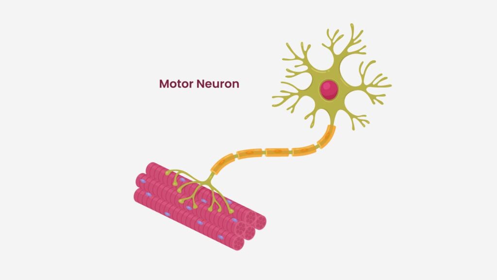
But what makes it move in the first place? Skeletal muscle is neurogenic, meaning it is innervated and that under physiological conditions it does not contract unless a nerve tells it to. A motor neuron fires, acetylcholine crosses the neuromuscular junction, the muscle fiber generates its own action potential, and only then does the muscle contract. Much of what skeletal muscle does is also voluntary, and those are two different properties.
Neurogenic describes where the activating signal originates. It says nothing about whether you chose to send it.
A withdrawal reflex can be organized in the spinal cord without any conscious decision to make it happen, and postural adjustments draw on spinal circuits together with brainstem and other pathways above them. Either way the contraction is still entirely neurogenic, because the motor neuron is still the final pathway into the muscle.
Losing that input does not simply switch the muscle off, either. Denervated muscle becomes flaccid and progressively atrophies, and individual denervated fibers can become hyperexcitable and produce small spontaneous discharges called fibrillation potentials. Those are not coordinated contractions, and they are not evidence that skeletal muscle has acquired a pacemaker of its own.
Cardiac Muscle Is Myogenic
Cardiac muscle is not neurogenic. It is myogenic, which means it contracts by itself. Pacemaker cells depolarize spontaneously, generate action potentials, and drive the contractile cells to contract, with no signal from the brain required.
That does not make the heart independent of the nervous system, because it is innervated by both sympathetic and parasympathetic fibers. Sympathetic stimulation acts on beta-1 adrenergic receptors, and it produces two effects worth naming separately.
A positive chronotropic effect raises heart rate, and a positive inotropic effect raises the force of each contraction. Beta-1 stimulation also speeds electrical conduction, a dromotropic effect, and speeds relaxation, a lusitropic effect.
Parasympathetic activity opposes some of that, with acetylcholine acting on muscarinic receptors mainly at the sinoatrial and atrioventricular nodes to slow the rate and slow conduction. Its direct effect on ventricular contractility is much smaller than the sympathetic effect, so the vagus is not simply the mirror image of everything above.
The transmitter released by the sympathetic nerves that supply the heart is mainly norepinephrine, released locally at the nerve ending. Epinephrine is the one that arrives through the circulation, secreted by the adrenal medulla when the sympathetic system activates it.
Cardiac beta-1 receptors respond to both, so during exercise or stress the two act together, but a neurotransmitter released at a nerve terminal and a hormone released into the blood are not the same thing.
So cardiac cells being myogenic and cardiac cells being influenced by the autonomic nervous system are both true at once. And when we say cardiac muscle, we are talking about the heart.
Smooth Muscle Is Involuntary and Largely Myogenic
Smooth muscle is involuntary and non-striated, and it lines the walls of hollow structures, including blood vessels, the gastrointestinal tract, the airways, and the urinary and reproductive tracts. Much of the visceral, single-unit smooth muscle is myogenic in the way cardiac muscle is, developing spontaneous or stretch-related activity, while multi-unit smooth muscle depends more heavily on autonomic input. Hormones and local mediators modulate both, and the single-unit and multi-unit division is more of a spectrum than a hard line.
Its contractile machinery is arranged differently too. Smooth muscle actin and myosin are not organized into sarcomeres, so force is transmitted through structures such as dense bodies rather than through the striated arrangement of skeletal and cardiac muscle.
And it regulates contraction differently, because rising intracellular calcium binds calmodulin, which activates myosin light-chain kinase, which phosphorylates myosin to allow cross-bridge cycling, rather than working through the calcium and troponin mechanism the other two types use.
Is Muscle an Organ? Each Skeletal Muscle Is One
There are somewhere north of 600 skeletal muscles in the human body, and each one of them is an organ. So you are walking around with at least that many organs. They are not 600 different kinds of organ, because they are built from the same tissues, but the biceps brachii and the soleus really are separate organs rather than two regions of one, and it is worth sitting with how many of them there are.
How Many Skeletal Muscles Are There?
Reputable sources give different counts. The National Cancer Institute’s anatomy material says more than 600. The Library of Congress notes that most sources give more than 650 named skeletal muscles, and that some counts reach around 840.
That spread is a counting convention problem rather than a gap in anatomical knowledge. It depends on whether the left and right members of a pair count once or twice, whether separate heads and bellies are counted independently, and how small or inconsistently present structures are handled. People genuinely vary as well, so no individual body has to match the atlas.
What Makes Something an Organ
An organ is a structure built from multiple tissue types working together toward a function, and a whole skeletal muscle meets that definition without any stretching.
It contains skeletal muscle tissue along with connective tissue, nerve tissue and vascular tissue, integrated into one coordinated unit. The National Cancer Institute’s teaching material states it plainly, that a whole skeletal muscle is considered an organ of the muscular system, made of skeletal muscle tissue together with connective, nerve and vascular tissue.
At the level of gross anatomy, an individual muscle such as the biceps brachii or the soleus is an organ, skeletal muscle tissue is a tissue, and the collection of those organs makes up the muscular system.
At the level of systems physiology, researchers also speak of skeletal muscle collectively as a metabolic and endocrine organ, because the distributed tissue mass carries out coordinated whole-body work in substrate metabolism, thermogenesis and secreted signaling. Both usages are legitimate. The second one is what the rest of this article is about.
Why Muscle Matters for Health, Disease and Aging
The obvious answers to why we study muscle are locomotion and posture, and they are real. But the bigger answer is health and disease, and then aging, which is closely related to disease.
That is the part that is underrated. Muscle influences health through more routes than one article can follow, and this one follows three. It secretes signaling molecules into the blood, it handles the glucose you eat, and it runs the intracellular signaling pathways that adaptation depends on. Thermogenesis, substrate oxidation, whole-body protein handling and physical function all belong on the longer list.
Muscle Is an Endocrine Organ That Secretes Myokines
Muscle tissue is an endocrine signaling tissue. It secretes myokines, which are signaling molecules released by muscle that act on muscle itself and on other tissues. This is the clearest reason to call muscle an organ in the physiological sense and not only the anatomical one, because a tissue that did nothing but produce force would have no need of an outbound signaling vocabulary at all.
Irisin
Irisin is one of them, and it is the most studied and the most contested. It was identified in 2012 as an exercise-responsive signal linked to PGC-1α, and in mouse and cell experiments, raising it drove a brown-fat-like program in white fat, including UCP1 expression and thermogenic remodeling, along with increases in energy expenditure. Those results are preclinical.
The human story then ran into two serious measurement problems. Many early studies of circulating irisin relied on commercial antibody assays, and in 2015 Albrecht and colleagues found that commonly used antibodies cross-reacted non-specifically and failed to identify the expected irisin band in human serum, which put a great many published concentrations in doubt.
In the same year Jedrychowski and colleagues took a different approach, using targeted mass spectrometry with labeled internal standards, and did detect circulating irisin in humans, at roughly 3.6 ng/mL in four sedentary people against 4.3 ng/mL in six people doing aerobic interval training.
Circulating irisin has been detected in human plasma by targeted mass spectrometry, which is far better evidence than the assays that came before it.
What remains unsettled is how reliably it can be measured routinely, how much exercise actually changes physiologically meaningful concentrations, and what those concentrations do once they are there. The browning and thermogenesis results are potential benefits in the preclinical sense, and none of them should be read as a demonstrated human outcome yet.
Interleukin 6 and the Anti-Inflammatory Effect of Exercise
Interleukin 6 is a cytokine, and it is usually described as pro-inflammatory. But contracting muscle releases it during exercise, with a larger response during prolonged work and when muscle glycogen is low, and in that context it takes part in a signaling environment that produces anti-inflammatory effects.
In a human infusion experiment that raised interleukin 6 to about 140 pg/mL, which is the range seen after strenuous prolonged exercise, it increased circulating interleukin 1 receptor antagonist and interleukin 10, both of them anti-inflammatory, without a corresponding rise in TNF-alpha.
Interleukin 1 receptor antagonist is the best characterized part of that response. It works by occupying the interleukin 1 receptor without activating it, so it blocks interleukin 1 beta from signaling through that receptor rather than producing an anti-inflammatory signal of its own.
Exercise can raise interleukin 4 as well, though less consistently than it raises interleukin 1 receptor antagonist and interleukin 10.
A cytokine’s effect depends on its source, its receptor and signaling mode, its timing, its concentration and the other cytokines around it rather than on a fixed label. So muscle-derived interleukin 6 is not an anti-inflammatory molecule in its own right, and a brief exercise-induced pulse of it is not biologically the same thing as chronically elevated interleukin 6.
The word hormetic fits the exercise response as a whole, a transient stress that produces an adaptive answer, and the muscle-derived interleukin 6 signal is one component of it.
Metabokines, and Why Lactate Is Not the Villain
Muscle also releases metabolites that act as signals rather than only as fuel, and these are sometimes called metabokines. The term is less settled than myokine, and the best example, lactate, is neither unique to muscle nor only a signal, since many tissues produce it and it is a genuine fuel as well.
Lactate Is Not the Same Thing as Lactic Acid
What accumulates in exercising muscle is lactate rather than lactic acid, and the two names are not interchangeable.
Lactic acid has a pKa of about 3.86, so at a blood pH of 7.4 roughly 99.97 percent of the pair exists in the dissociated lactate form, and even at an intracellular pH of 6.5 it is still more than 99.7 percent dissociated.
The conjugate acid does not cease to exist, since equilibrium favoring the anion is not the same as the acid being absent. Essentially all of the pair is lactate, so lactate accumulation is the accurate description of what happens in a working muscle.
Producing lactate is also not the source of the acidity that accompanies hard exercise. The reaction catalyzed by lactate dehydrogenase consumes a proton as pyruvate becomes lactate, which is a large part of why exercise biochemistry moved away from the lactic acid explanation of acidosis.
Does Lactate Cause Soreness or Fatigue?
Lactate does not cause soreness, and it is not a primary direct cause of fatigue either. Delayed onset muscle soreness appears many hours after unfamiliar work, especially eccentric loading, and it does not track lactate at all.
Exercise can produce high lactate with little soreness, eccentric work can produce marked soreness without much lactate, and lactate has returned toward baseline long before soreness peaks. Soreness is better understood as a mechanically initiated process, with connective tissue disturbance, inflammatory signaling and sensitization of pain receptors among the proposed contributors. The precise mechanism is still unsettled, and soreness can appear without the amount of fiber damage the traditional damage-first account assumes.
Fatigue is multifactorial. Inorganic phosphate accumulation, potassium redistribution, impaired action potential propagation, calcium handling and central factors all contribute, and lactate is not the villain in that story either.
There is evidence pointing the other way, inside narrow conditions. In isolated rat muscle made poorly excitable by high extracellular potassium, lactate improved excitability by inhibiting the ClC-1 chloride conductance, and force largely recovered.
That is a real mechanism. It is also an isolated preparation under artificial conditions, so it does not establish that circulating lactate acts as an anti-fatigue agent in exercising humans.
Lactate, BDNF and Brain Health
Lactate matters because the brain can import and oxidize it efficiently, as can other tissues including the heart, and cerebral uptake rises when circulating lactate rises, including during exercise. Glucose is still the brain’s predominant circulating fuel at rest. Lactate also appears to move BDNF, brain derived neurotrophic factor, which supports neurogenesis and brain health generally. Which form of it moves matters, because the precursor and the mature protein act on different receptors and can pull in opposite directions.
That may be one of the links explaining why exercise does so much for cognitive function, executive function and brain health. Potentially because lactate activates BDNF. There are a lot of other pathways as well, but that is a big one.
The evidence sits at different strengths depending on which step of it you are looking at. In mice, lactate has been proposed to engage a SIRT1 to PGC-1α to FNDC5 to BDNF pathway in the hippocampus, with associated improvements on learning and memory tasks. In humans the work has moved beyond association.
A 2025 infusion study in 18 healthy young adults raised plasma lactate to about 5.9 mmol/L and saw plasma pro-BDNF rise by roughly 55 to 68 percent during recovery, though mature BDNF did not significantly increase, and a separate infusion experiment during exercise did amplify the mature BDNF response.
What none of that shows is that exercise-derived lactate raises BDNF in the human hippocampus, or that this pathway is the reason exercise helps cognition. However, what is clear is that exercise does help cognition, and there are multiple plausible pathways that may contribute.
Muscle Is the Primary Postprandial Glucose Sink
When you eat, the carbohydrate you have just ingested has to go somewhere.
How Little Glucose Is Actually in Your Blood
Consider how tightly blood glucose has to be regulated. You have roughly 5 liters of blood. Treat 100 milligrams per deciliter as a whole-blood concentration, which is a round number chosen for the arithmetic, and that works out to about 5 grams of glucose in your entire circulation.
Roughly a teaspoon. That is not much. It is a teaching approximation rather than a measurement, because clinical results are reported as plasma glucose, and plasma and whole blood do not carry glucose at identical concentrations once hematocrit and water distribution are accounted for.
Measured fasting, 100 mg/dL is the lower boundary of the range the American Diabetes Association calls impaired fasting glucose, which runs from 100 to 125 mg/dL. Normal fasting plasma glucose is below 100, so the round number chosen for the arithmetic is not a comfortably normal one. Measured after a meal it means something different, because glucose is supposed to rise after you eat.
What Half a Teaspoon of Blood Glucose Actually Does
Now halve the amount in circulation. Two and a half grams of glucose in 5 liters of blood works out to about 50 mg/dL, or 2.8 mmol/L, and that is clinically significant hypoglycemia, a range in which cognition and brain function are often impaired.
The deterioration is graded rather than switched. The American Diabetes Association classifies glucose below 70 but at or above 54 mg/dL as level 1 hypoglycemia, and below 54 mg/dL as level 2, where neuroglycopenia and meaningful cognitive impairment become a real concern. Level 3 is defined by severe impairment requiring another person’s assistance, deliberately with no glucose number attached to it at all, because there is no concentration at which a person reliably becomes comatose.
The measured threshold in healthy people sits a little below the clinical one, with cognitive function generally holding up until glucose falls under about 50 mg/dL, or 2.8 mmol/L. Severe hypoglycemia can certainly progress through confusion and abnormal behavior to seizure and loss of consciousness, and it does so in some people at values that others tolerate.
Why the Hypoglycemia Threshold Varies From Person to Person
Personal history is a large part of why. Repeated hypoglycemia produces hypoglycemia-associated autonomic failure, which shifts the warning symptoms down to lower glucose concentrations, so somebody with recurrent lows can look functional at a number that would visibly affect somebody else.
It runs the other way too. Somebody accustomed to chronic hyperglycemia can experience hypoglycemic symptoms during a rapid fall while their glucose is still inside the normal range.
What 200 mg/dL Does and Does Not Diagnose
Now double the original number instead, adding another teaspoon of sugar to the blood, and you are at about 200 mg/dL. It is diagnostic of diabetes at two hours during a standardized 75 gram oral glucose tolerance test, or as a random plasma glucose in somebody with classic hyperglycemic symptoms.
The other criteria are a fasting plasma glucose at or above 126 mg/dL, and an HbA1c at or above 6.5 percent. And in the absence of unequivocal symptoms, the diagnosis requires two abnormal results rather than one.
A one-off peak near 200 mg/dL after an ordinary meal is a different situation. When people classified as normal by fasting glucose, an oral glucose tolerance test and HbA1c wore continuous glucose monitors, those with the most variable patterns spent up to 15 percent of the recording above 140 mg/dL, which the investigators classed as a prediabetic range, and about 2 percent of it above 200 mg/dL.
Those are that study’s categories rather than diagnostic ones, and the American Diabetes Association is explicit that the evidence does not support using continuous monitoring to screen for or diagnose either prediabetes or diabetes.
So a high reading on a monitor is a reason to get properly tested rather than a diagnosis in itself. The person may simply have just eaten, or may have poor glucose control that falls short of diabetes.
Glycation, HbA1c and the Cost of Sustained High Glucose
What sustained high glucose does bring with it is glycation. Glycation is the spontaneous, non-enzymatic reaction of sugars such as glucose with amino groups on proteins, and further rearrangement and cross-linking can turn some of those products into advanced glycation end products, or AGEs.
It is not the same thing as glycosylation, which is the regulated, enzyme-controlled attachment of sugars that cells carry out on purpose.
You have already met a glycation product if you have ever had an HbA1c test, because HbA1c is what forms when glucose glycates hemoglobin over the red cell’s lifespan, which is also why it integrates your glucose exposure over roughly the previous 8 to 12 weeks instead of reporting a single moment. That window holds as long as red cell turnover and hemoglobin are reasonably normal, and altered turnover, hemoglobin variants, recent blood loss or transfusion and some kidney and blood disorders can pull HbA1c away from true average glucose.
What Happens When You Do Not Have Enough Muscle
So where does the glucose go? Muscle stores a lot of it as glycogen. That is why I describe muscle as the primary postprandial glucose sink, which is a label I made up for the job rather than established terminology.
How Much of a Meal Muscle Actually Takes Up
Under experimental hyperinsulinemic clamp conditions, skeletal muscle accounts for the large majority of insulin-stimulated glucose disposal, which is where its reputation comes from. The figure usually quoted is 70 to 90 percent, and that range is wide because the studies behind it were not all measuring the same thing. A review of the clamp literature puts skeletal muscle at 70 to 75 percent of insulin-stimulated glucose disposal, while a study that catheterized the leg directly estimated that about 90 percent of the infused glucose was disposed of by peripheral tissue, which in the leg is predominantly muscle. The share is not fixed within a population either, since one study estimated about 83 percent in non-obese people against about 64 percent in obese people. The share falls as insulin resistance rises.
A real meal is more distributed, because oral carbohydrate reaches the liver through the portal circulation before the rest of the body sees it. In one five-hour mixed-meal study in 11 men, skeletal muscle took up about 23 grams, roughly 30 percent of the ingested starch, while the splanchnic bed took about 29 grams, or 39 percent. Only about 60 percent of that starch had been absorbed inside the measurement window, so these are the results of one experiment rather than a universal partition of every meal.
So muscle is the major insulin-sensitive site of glucose disposal, and the clamp figure and the meal figure answer two different questions.
Where the Glucose Goes When Muscle Cannot Take It
When there is not much muscle to store the glucose in, or when the muscle that is there is insulin resistant, more of the glucose stays in circulation for longer, and other tissues, including the liver, take on more of the job. The liver can oxidize it, store it as glycogen, or convert carbohydrate-derived acetyl-CoA into fatty acids through de novo lipogenesis, running glucose to pyruvate, on to citrate, and out into fat.
Measured against the liver’s own triglyceride, de novo lipogenesis is substantial and it climbs steeply with insulin resistance.
Tracer work estimates its contribution to newly made intrahepatic triglyceride palmitate at about 11 percent in lean people, 19 percent in people who are obese with normal liver fat, and 38 percent in people with fatty liver, with an earlier study putting the fatty liver figure at 26 percent. That is what the pathway contributed while it was being measured, rather than the origin of the fat already sitting there.
Measured as a share of the carbohydrate you just ate, it is a much smaller number, because on an energy-balanced mixed diet converting dietary carbohydrate into fat is a minor fate for it.
So the more common consequence of poor muscle glucose uptake is larger and longer postprandial hyperglycemia, rather than the meal being converted into body fat on the spot.
Does More Muscle Prevent Type 2 Diabetes?
More muscle tracks with better glucose regulation. In an NHANES analysis of 13,644 adults, each 10 percent increase in skeletal muscle index was associated with about an 11 percent lower HOMA-IR and a 12 percent lower prevalence of the study’s combined pre-diabetic or diabetic outcome, and the authors were explicit that this is observational and needs prospective work behind it.
Muscle Quantity Is Not the Same as Muscle Quality
Muscle quantity is also not the same variable as muscle quality. Two people with the same muscle mass can differ substantially in intramuscular fat, mitochondrial phenotype, capillary supply and insulin-stimulated glucose transport.
The stronger causal evidence is for training. Resistance training improves insulin sensitivity at the same time as it increases size, and in one six-week study in ten overweight men it raised an oral-test-derived insulin sensitivity index by about 16 percent alongside a 10.3 percent increase in muscle thickness.
Ten men is a small study and the spread on that insulin sensitivity figure was wider than the figure itself, so treat it as an indication rather than a measurement. And because training changes glucose transport, signaling, perfusion and enzyme capacity simultaneously with size, the result does not isolate the added mass as the cause.
Maintaining adequate, trained, insulin-sensitive muscle supports glucose disposal, and it is associated with a lower prevalence of prediabetes and diabetes together. Age-related loss of metabolically active muscle can contribute to a reduced capacity to clear a meal, particularly where it arrives alongside falling insulin sensitivity, rising adiposity and less physical activity.
What the evidence supports most strongly is trained, insulin-sensitive muscle, which is a good reason to keep yours and to keep working it.
The Signaling Pathways That Make Muscle Adapt
The other thing muscle does is signal internally. This is intracellular signaling, and it is how the tissue adapts to what you ask of it.
AMPK is a cellular energy sensor. Its activity rises when AMP and ADP rise relative to ATP, and in muscle it shifts metabolism toward generating ATP, increasing glucose transport and fatty acid oxidation while restraining the processes that spend energy. It senses energy status rather than exercise as such.
PGC-1α is a transcriptional coactivator rather than a sensor in its own right. It integrates the contraction-responsive calcium and kinase signals and coactivates transcriptional programs for mitochondrial biogenesis, and it is a major contributor to the rise in mitochondrial content that training produces rather than the whole explanation for it, since several partly redundant pathways feed the same adaptation.
NRF2 is a redox-responsive and electrophile-responsive transcription factor, normally held in check by KEAP1, and when oxidative or electrophilic stress modifies that control system, NRF2 is stabilized and switches on antioxidant response element genes covering antioxidant defense, glutathione and NADPH metabolism, and detoxification.
mTOR, and in this context specifically mTOR complex 1, integrates amino acid availability, insulin and growth factor signals, cellular energy status and mechanical loading, and drives the protein synthesis and ribosome biogenesis that mediate hypertrophy. There are many more of these than a single article can list.
Muscle, Performance and How You Feel About Your Body
There are other reasons to understand muscle. Athletic performance is one. How do you get stronger, how do you develop more force, what do the force-velocity relationships look like, and what raises neural drive.
Then there are the psychological factors. Physical appearance, how you look and how you feel about yourself. Body composition, quality of life, and the ability to avoid injury. All of it feeds into overall well-being, and into the psychology and the sociology around it.
Muscle Is an Underrated Organ
Muscle is a very important organ, and an underrated one. It is often not understood to be an organ in and of itself, one that influences the rest of the body systemically.
Each skeletal muscle meets the anatomical definition of an organ. Collectively, skeletal muscle meets the physiological one, through the three routes covered here.
It secretes myokines and metabokines that act on distant tissues, including the brain. It is the major insulin-sensitive site of glucose disposal, which is why muscle keeps appearing in the metabolic literature at all. And it runs the signaling pathways that let it remodel itself in response to what you ask of it.
Treating muscle as the tissue that moves you around undersells it considerably.
Tyler W. LeBaron, MSc., PhD.
Tyler W. LeBaron, MSc, PhD is a is a researcher and educator who translates complex science into practical insight on health, performance, and human potential. He is the Founder and Executive Director of the Molecular Hydrogen Institute (a science-based 501(c)3 nonprofit) and an adjunct professor of exercise physiology and chemistry at Southern Utah University. Tyler is known for evidence-based, engaging presentations that challenge assumptions, clarify emerging science, and inspire high-performing individuals and organizations. He has 80+ peer-reviewed publications, 2,000+ citations, top 0.5% recognition in oxidative stress, and has delivered invited talks on six continents.
Disclaimer: This blog is for general informational purposes only and does not constitute the practice of medicine, nursing or other professional health care services, including the giving of medical advice, and no doctor/patient relationship is formed. The use of information on this blog or materials linked from this blog is at the user’s own risk. The content of this blog is not intended to be a substitute for professional medical advice, diagnosis, or treatment. Users should not disregard, or delay in obtaining, medical advice for any medical condition they may have, and should seek the assistance of their health care professionals for any such conditions.
You may also like these…